Computer vision · Life sciences
Laboratory & microscopy image analysis
Counting, measuring, and classifying — hours of microscope work turned into minutes.
First working version in 6–10 weeks
The problem
Labs generate far more images than people can analyze: cell cultures, organisms, growth structures, samples. Manual analysis is a bottleneck that limits throughput, and results vary between analysts.
Off-the-shelf tools rarely fit specialized assays; the winning system speaks your protocol — magnification, staining, growth stages, edge cases — and integrates with how your lab already records results.
Our approach
Assay and image audit
We review your imaging setup, sample variability, and what you currently measure by hand.
Segmentation and measurement models
Trained on your images, evaluated against expert annotations, with disagreement analysis where experts differ.
Quantification pipeline
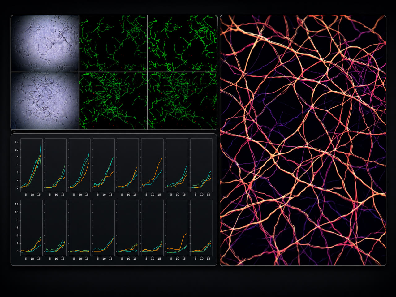
From raw images to counts, sizes, morphology, growth curves, and statistics — exported to your LIMS or notebooks.
Review workflow
Analysts verify and correct edge cases; every correction improves the model.
Automation hooks
Where useful, connection to acquisition hardware, plate readers, or bioreactor monitoring for closed-loop experiments.
Reference architecture
- Image ingestion from microscopes, scanners, or acquisition rigs
- Segmentation and classification models tuned to your assay
- Measurement and statistics pipeline with calibrated units
- Analyst review interface with correction capture
- Export to LIMS, notebooks, or analysis environments
What you get
- A validated analysis pipeline for your specific assay
- Agreement metrics against your experts' annotations
- Throughput your manual process cannot reach
- Reproducible, analyst-independent results
Proof. GI engineered AI-driven biological image analysis in the technology of Eternal's space division — winner of NASA's Deep Space Food Challenge.
Turn microscope hours into minutes.
A 30-minute call is enough to tell you whether this is feasible on your data.